15+ Brain Tumor Images Mri UK. Brain tumor segmentation plays an important role in medical image processing. Magnetic resonance imaging (mri) is an advanced imaging technique that is used to observe a variety of diseases and parts of the body.neural networks can analyze these images individually (as a radiologist would) or combine them into a single 3d volume to make predictions.
New Standardized Brain Tumor MRI Recommendations Announced ... from www.itnonline.com Film mri ( magnetic resonance imaging ) of brain ( stroke , brain tumor , cerebral infarction , intracerebral hemorrhage ) ( medical , health care , science background ) ( cross section of brain )fil. Diagnostic tools include computed tomography (ct or cat scan) and magnetic resonance imaging (mri). The following instructions will first describe the methods for image analysis through filtering and cleaning up the mri scan, through binarizing.
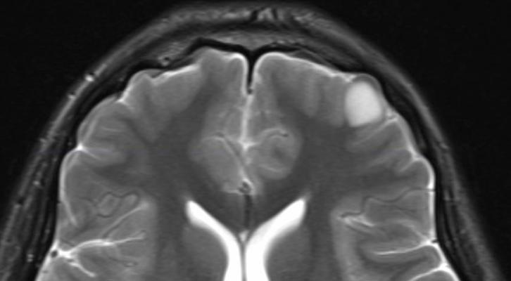
Once mri shows that there is a tumor in the brain, the most common.
Show brain tumor at right parietal lobe of cerebrum. A ct scan or an mri report usually detects a benign brain tumor without difficulty. This program is designed to originally work with tumor detection in brain mri scans, but it can also be used for cancer diagnostics in other organ scans as well. There are different brain tumor detection and segmentation methods to detect and segment a brain tumor from mri images.
Komentar
Posting Komentar